The Ankle - Syndesmosis (High) Ankle Injury
High ankle sprains are complex and require skilled input to rehabilitate effectively and return to sport.

Sprained your ankle? See why we're recommending hydrotherapy for higher grade ankle injuries.
Ankle sprains are the most common musculoskeletal injury worldwide, so there is a good chance most of us will experience at least one ankle injury during our lifetime. Most of these will impact the outside of the ankle, called a lateral ligament sprain. More complex sprains of the ankle can involve a structure called the syndesmosis, and these can be difficult to diagnose and more challenging to rehabilitate. Read on to learn a little more about syndesmosis injuries and how they get the name ‘high ankle sprain’.
A little bit of anatomy
A syndesmosis is defined as a fibrous joint between two adjacent bones, held together by a strong membrane and ligaments. The tibiofibular syndesmosis sits just above the ankle joint (thus why it's referred to as a 'high' ankle sprain) and consists of...yep you may have guessed it...the tibia on the inside and the fibula on the outside. The main ligaments holding the syndesmosis together are the inferior tibiofibular ligaments (there’s an anterior one and a posterior one) and the lower fibers of the interosseous membrane. On the inside of the ankle is the super strong deltoid ligament, while on the outside there are a few ligaments, namely the anterior talofibular ligament, calcaneofibular ligament and the posterior talofibular ligament. Collectively these ligaments, along with the muscles in this region, provide stability to the ankle joint and allow the foot to adjust to the surfaces underneath and produce power for locomotion.
Who gets it?
Syndesmosis injuries represent up to 18% of all ankle sprains in the general population but spike to 32% in athletic populations. They often involve tears in the ligaments connecting the tibia and fibula, leading to instability. These injuries usually occur when the foot forcefully rotates outward while the ankle is bent upward and the foot turned inward, potentially damaging inner ligaments. Factors like excessive foot bending or twisting can contribute and they're sometimes part of a more complex fracture pattern called Maisonneuve fracture.
Diagnosing high ankle sprain
Patients with syndesmosis ligament injuries often report a history of ankle trauma. Common symptoms include pain in the front part of the ankle during weight-bearing, worsened by rotating the foot outward and pulling it upward. Signs include feelings of ankle instability, tenderness, swelling, and difficulty bending the foot upward, with recurring swelling in the joint. Chronic cases may involve pain in the front and outer part of the ankle, frequent swelling, instability/giving way, and sometimes stiffness.
Diagnosis involves comparing the injured ankle to the unaffected one and performing specific physical
tests like external rotation, squeeze test, Cotton, and fibular translation tests.
These tests help detect pain, instability, or abnormal movement patterns and help determine whether a syndesmosis injury is present or whether symptoms are caused by something else, such as a lateral ankle sprain, ankle fracture or tendon injury.
Do I need a scan?
X-rays are usually indicated when a high ankle sprain is suspected and can be ordered by your physiotherapist. Syndesmosis injuries require significant force, and fractures need to be considered as part of the differential diagnosis. Xray can also assist with determining if surgical intervention is required or if the injury can be managed conservatively.
In some circumstances, other imaging techniques can be beneficial. Ultrasound and weight bearing CT scans can evaluate the ankle’s stability, comparing both sides for subtle differences in bone position under stress. MRI, while being highly sensitive for certain ankle injuries, may not clearly show syndesmosis instability without additional functional tests.
Treatment
When managing syndesmosis injuries, treatment depends on ankle stability. For stable cases with bones well-aligned the PEACE and LOVE principle is used.
An immobilizing brace can be used to limit rotation, and activity is often restricted for four to six weeks.
Physiotherapy is crucial in the management and rehabilitation of high ankle injuries. Physiotherapy aims to optimize healing by restoring joint function, reducing pain, improving mobility, and enhancing ankle stability, helping patients regain activity levels and reducing long-term complications like chronic instability or joint degeneration.
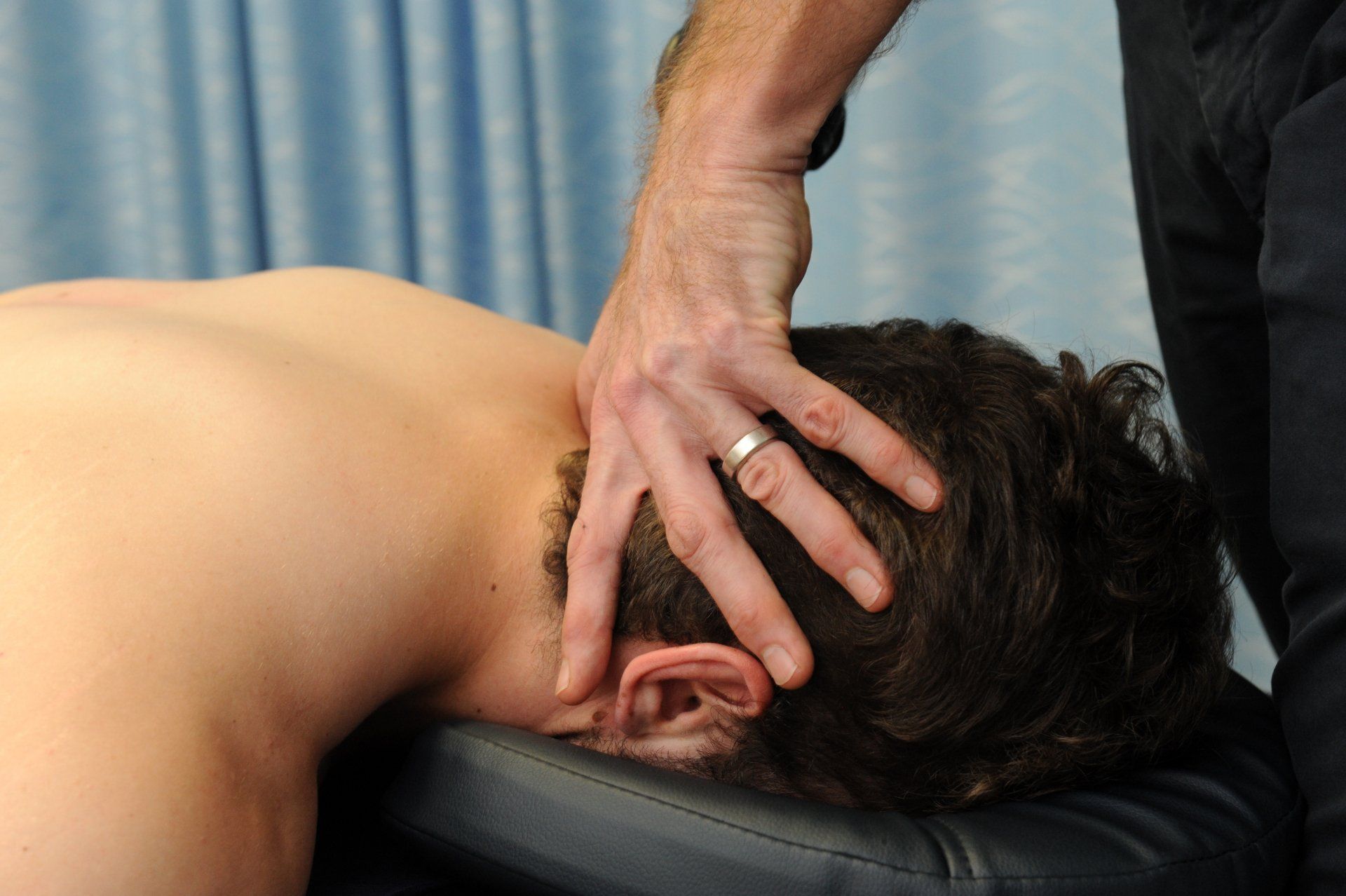
Early phase rehabilitation utilises manual and soft tissue therapies to reduce pain and swelling, and restore joint mobility. Dry needling can be beneficial to redruce muscle tension and pain. Hydrotherapy is a great exercise medium in the early stages, offering gentle resistance for improved ankle mobilization and strengthening, and reducing swelling via increased hydrostatic pressure.
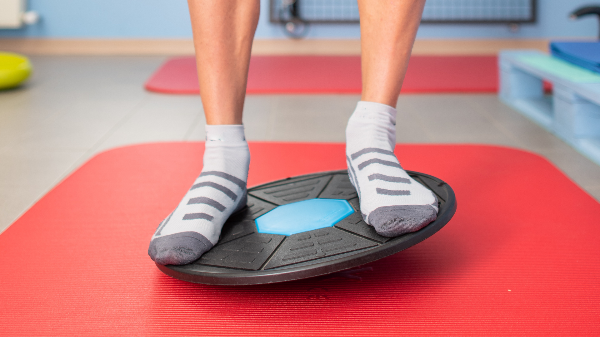
Graded therapeutic exercise programs, tailored by physiotherapists, start with basic
exercises for ankle mobility, proprioception and balance.
These are progressed to include functional exercises mimicking daily or sports activities, improving strength, agility and power, and thereby reducing the risk of a recurrent injury.
Will I need surgery?
Surgery is indicated for acute first-time injuries if disruption of the joint space is significant (high grade) and/or there is a fracture present that requires surgical fixation. Surgery may also be considered in some chronic and recurrent cases where conservative management has failed. Surgery usually entails screw fixation to stabilize the syndesmosis, followed by a period of limited weight bearing and rehabilitation. In many cases the screw is removed later when healing is complete.
How long’s it going to take?
The outlook for syndesmosis injuries depends on whether the injury is recent (acute) or persisting (chronic), and if it’s a first-time injury or recurrence of a previous ankle injury. Low to moderate grade acute injuries can heal well if treated promptly and diligently, allowing most patients to return to sports in approximately 6-12 weeks.
Chronic cases, where symptoms have been present for over six months, and/or recurrent injuries, often indicates more complex issues.
These presentations may require surgical intervention to stabilize ligaments, resulting in a prolonged recovery and rehabilitation period lasting 4-6 months.
The Take Home
Ankle syndesmosis injuries vary in severity and require careful diagnosis and treatment. They affect the ligaments between the tibia and fibula bones that are crucial for ankle stability. Athletes and dancers are especially prone to these injuries due to intense physical activity. Diagnosis involves comparing both ankles and using various clinical and radiological tests. Treatment ranges from conservative management for stable injuries to surgery for more complex cases. Physiotherapy plays a vital role, focusing on early restoration of ankle movement and strength, functional rehabilitation, and return to sport. Early intervention is crucial for better outcomes, ensuring a quicker return to normal activities.
Injure your ankle and not sure what to do? Give us a call now.
At Movement for Life Physiotherapy, we can assess and diagnose the cause of your ankle pain and let you know whether you have a high ankle sprain, lateral ankle sprain, tendon injury, or if there is something else going on. With a clear diagnosis and tailored management plan, we'll help get you back to the things you love sooner.
Call us now on
Sources
- Calder JD, Bamford R, Petrie A, McCollum GA (2016) Stable versus unstable grade II high ankle sprains: a prospective study predicting the need for surgical stabilization and time to return to sports. Arthroscopy 32(4):634–642
- Hannon, C. P., Weber, D. C., & Golano, P. (2013). Treatment of chronic syndesmotic injury: A systematic review and meta-analysis. Foot & Ankle International, 34(4), 602-610. Retrieved from https://www.researchgate.net/profile/Charles-Hannon/publication/236339290_Treatment_of_Chronic_Syndesmotic_Injury_A_Systematic_Review_and_Meta-Analysis/links/55b8edfb08aed621de08046a/Treatment-of-Chronic-Syndesmotic-Injury-A-Systematic-Review-and-Meta-Analysis.pdf
- Hermans, J. J., Beumer, A., de Jong, T. A., & Kleinrensink, G. J. (2010). Anatomy of the distal tibiofibular syndesmosis in adults: a pictorial essay with a multimodality approach. Journal of anatomy, 217(6), 633–645. https://doi.org/10.1111/j.1469-7580.2010.01302.x
- Rellensmann, K., Behzadi, C., Usseglio, J. et al. Acute, isolated and unstable syndesmotic injuries are frequently associated with intra-articular pathologies. Knee Surg Sports Traumatol Arthrosc 29, 1516–1522 (2021). https://doi.org/10.1007/s00167-020-06141-y
- Saad, T. A. (2020). Comparative study between syndesmotic and suprasyndesmotic technique in syndesmotic ankle injury. Journal of Arthroscopy and Joint Surgery, 7(2), 91-97.
- https://www.orthobullets.com/foot-and-ankle/7029/high-ankle-sprain-and-syndesmosis-injury


