The Knee - Pes Anserine Tendino-bursitis
Pes Anserine bursitis is a common cause of medial knee pain in females with diabetes.
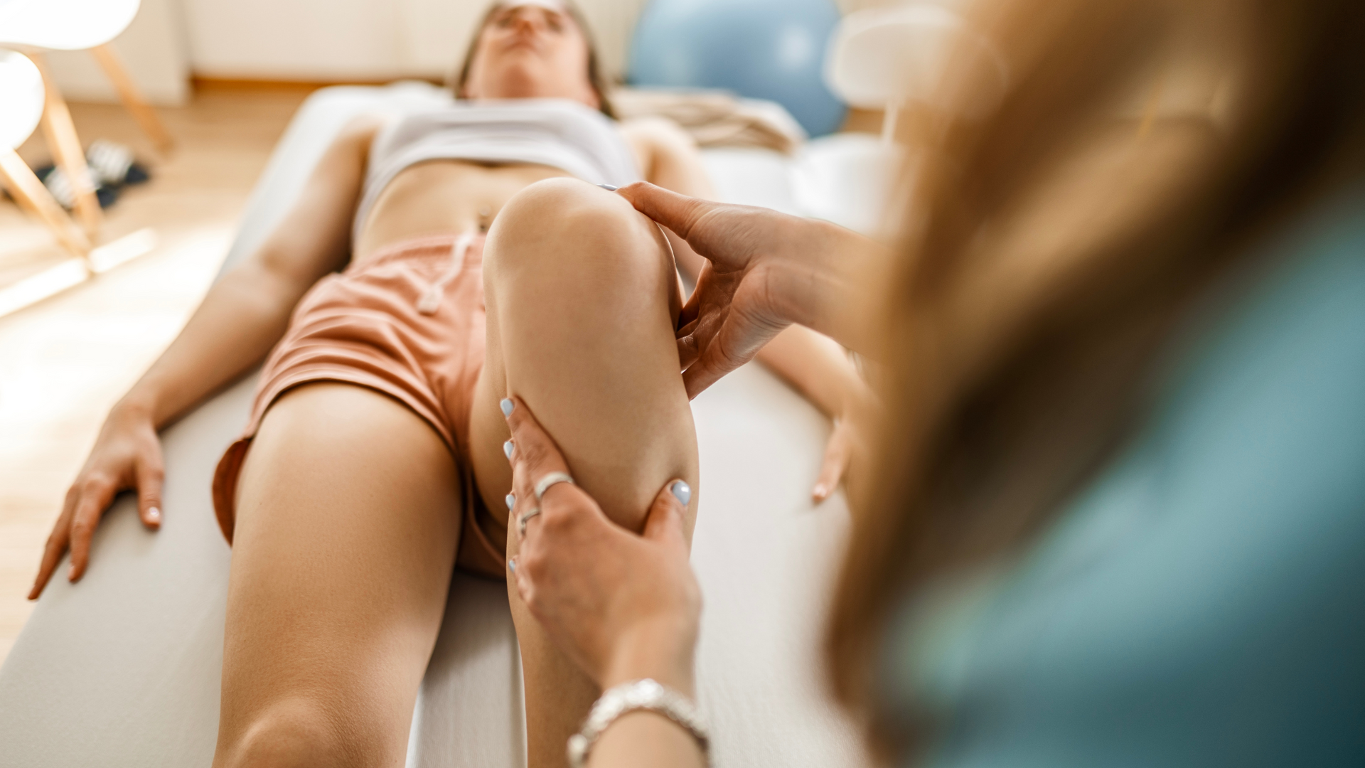
Many of us when we have knee pain immediately think of the joint. But there are lots of different structures around the knee, including muscles, ligaments, tendons and bursa that can all contribute to knee pain. Bursa and their associated tendon(s) are often overlooked as a source of irritation and a cause of pain. These small fluid-filled sacs are highly innervated (ie. they have lots of nerves) and susceptible to repetitive trauma and compression. The pes anserine bursa is one such structure, that can produce medial knee pain. As inflammation usually occurs in combination with tendinopathy, it is referred to as pes anserine tendino-bursitis (PATB).
A little bit of anatomy
Bursae are important structures that act as a buffer between a tendon and the bony surface underneath. Think of them like a thin pillow of fluid that disperses compressive forces and reduces friction between moving parts.
The pes anserine bursa is located on the medial (inside) aspect of the knee where three important muscles converge – semitendinosis (one of the hamstring muscles), gracilis (an adductor or groin muscle) and sartorius (a hip and knee flexor and rotator).
The convergence of these three muscles is known as the pes anserine, named for its resemblance to a gooses foot (in Latin, pes means “foot” and anserine “goose-like’). Just underneath the pes anserine is the bursa, which separates the tendon structure from the bone underneath.
The muscles that form the pes anserine come from three different compartments of the leg – the anterior, medial and posterior compartments. Together they provide hip stability and protect the knee against the rotation or valgus (knock-knees) stress. Changes to knee joint alignment, a sudden increase or repetitive use of these muscles, or sustained compression on the medial aspect of the knee, can irritate the tendon and bursa resulting in PATB.
Who gets it?
Overall, the incidence of PATB in an adult population is reported by various studies to be between 2.5 to 10%. PATB is more common in older females, particularly those with valgus knee deformity. Females naturally are more likely to have a wider pelvis (for childbirth), resulting in a greater angle at the knee joint (known as the Q angle). This places greater stress on the medial aspect of the knee and the structures supporting movement.
Diabetes is a known risk factor for PATB, while osteoarthritis and obesity are considered additional risk factors, though their role in the pathophysiology of disease is not yet understood.
Causes of PATB
One of the more common reasons PATB develops is overuse or repetitive trauma. Exposure of the medial knee joint line to repetitive load stresses the tendon. Without adequate recovery periods this can cause pathological changes to the tendon complex, including the bursa.
Activities such as running on hard surfaces, walking uphill and /or downhill regularly, repetitive squatting, or sports that require running and change of direction can all contribute to the development of PATB.
Other contributing factors to PATB can include:
- Injury or direct trauma- a fall or direct blow to the medial side of the knee can cause localised trauma that proceeds to PATB.
- Biomechanical and/or developmental changes - malalignment of the knee joint producing a greater Q angle, or muscle imbalance resulting in reduced knee joint motion control can increase load and stress on the medial aspect of the knee.
- Poor footwear – while empirical evidence linking foot posture and control to PATB is lacking, altered foot posture and control is known to influence knee joint biomechanics. Anecdotally this has contributed to symptoms of PATB.
Diagnosing PATB
PATB is a clinical diagnosis that can be made by a physiotherapist.
People with PATB will often describe difficulty ascending and descending stairs, pain when walking for longer distances, pain when pivoting on the affected leg, and difficulty getting up from a sitting or lying position. Swelling, pain and tenderness on palpation are other indicators of pes anserinus tendino-bursitis.
PATB shares symptoms with a number of other conditions such as medial meniscal injury, medial collateral ligament injury and patellofemoral pain syndrome, and often co-exists with knee joint osteoarthritis, so make sure you get your knee pain assessed by a qualified health professional.
Do I need a scan?
Medical imaging is not usually required to diagnose PATB. In recalcitrant cases, or where other pathology is being considered, ultrasound or MRI can be useful.
Treatment and management of PATB
PATB can usually be effectively managed with a course of physiotherapy. Initial management should be aimed at education and goal setting. Identifying modifiable risk factors and addressing these will assist in reducing symptoms and pain - things like reducing walking speed or distance, altering exercise type and cross training.
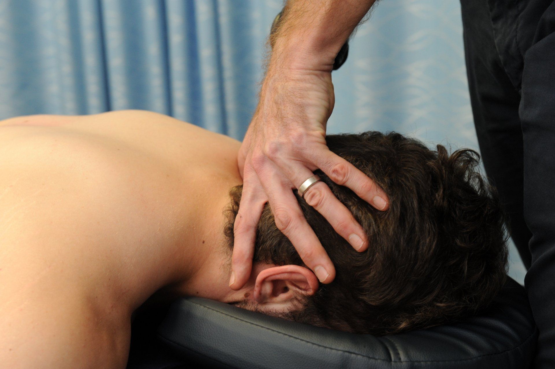
Early soft tissue and manual therapy in the first 2-3 weeks can be helpful to settle pain, particularly in the presence of co-morbidities such as knee osteoarthritis.
Exercise therapy is an important component in the treatment and management of PATB. Given the right load/recovery environment, tendon will respond by increasing tensile strength and resilience. If all load is removed, then the tendon simply adapts to this new environment by getting thinner and weaker. Not ideal!
A tailored, progressive, therapeutic exercise program will help keep the tendon healthy while pain subsides, and have the tendon primed and ready to receive load when able. This is crucial to the individual getting back to activity and pursuing their original goals.
Wholistic client-centred treatment will include advice on braces and supports and foot orthotics where indicated, and support for lifestyle modifications such as weight loss.
Can I get an injection?
Ultrasound guided intra-bursal corticosteroid injections have been shown to have good short-term effects on pain in about 70% of cases. However, studies comparing the long-term outcomes of CSI versus physiotherapy delivered exercise and education show no significant difference between groups for pain and function at 8 weeks.
CSI is known to detrimentally effect tendon structure, causing breakdown of the tendon matrix and reducing is load bearing capacity. It does not circumvent the need to address modifiable risk factors or to undertake therapeutic exercise. Thus, CSI is not recommended other than in recalcitrant cases.
How long’s it going to take?
The earlier PATB is diagnosed and intervention commenced, the better outcome. Recovery depends on the number of non-modifiable and modifiable risk factors present, the duration and severity of symptoms, and compliance with education and exercise program. In most instances symptoms can be settled within 3-6 weeks, with graded return to full activity taking 3-6 months.
Take home Advice
PATB is a common cause of medial knee pain, particularly in older diabetic females. It can be well managed by your physiotherapist with a wholistic approach covering load management, therapeutic exercise and education. Symptoms can take up to 6 weeks to settle, with most people making a full recovery and returning to pre-injury activity with no further symptoms.
Got knee pain and want to get it sorted? Give us a call.
At Movement for Life Physiotherapy, we can assess, diagnose, and treat your knee pain. We provide education and evidence-based advice coupled with a comprehensive treatment plan to help get you back to the things you love doing sooner.
Give us a call now or click on BOOK AN APPOINTMENT to book online.
References
- Alvarez-Nemegyei, J., 2007. Risk factors for pes anserinus tendinitis/bursitis syndrome: a case control study. JCR: Journal of Clinical Rheumatology, 13(2), pp.63-65.
- Curtis, B. R., Huang, B. K., Pathria, M. N., Resnick, D. L., & Smitaman, E. (2019). Pes anserinus: anatomy and pathology of native and harvested tendons. American Journal of Roentgenology, 213(5),
1107-1116. - Farsad, F., Moghimi, J., Mirmohammadkhani, M., Gholami, E., & Moazeni, M. (2023). The Effect of Modifying the Sitting and Getting up Method on Pain Intensity in Patients with Pes anserine tendinitis bursitis: A Randomized Clinical Trial Study.
- Gouda, W., Abbas, A. S., Abdel-Aziz, T. M., Shoaeir, M. Z., Ahmed, W., Moshrif, A., ... & Kamal, M. (2023). Comparing the Efficacy of Local Corticosteroid Injection, Platelet‐Rich Plasma, and Extracorporeal Shockwave Therapy in the Treatment of Pes Anserine Bursitis: A Prospective, Randomized, Comparative Study. Advances in Orthopedics, 2023(1),
5545520. - Helfenstein Jr, M. and Kuromoto, J., 2010. Anserine syndrome. Revista brasileira de reumatologia, 50, pp.313-327.
- Patil, A., Dass, B., Hotwani, R., Kulkarni, C. A., Naqvi, W. M., & Wadhokar, O. C. The Bio-mechanical correction exercises in pes anserine bursitis. Knee, 2, 5.
- Saggini, R., Di Stefano, A., Dodaj, I., Scarcello, L., & Bellomo, R. G. (2015). Pes anserine bursitis in symptomatic osteoarthritis patients: a mesotherapy treatment study. The Journal of Alternative and Complementary Medicine, 21(8), 480-484.
- Sarifakioglu, B., Afsar, S.I., Yalbuzdag, S.A., Ustaömer, K. and Bayramoğlu, M., 2016. Comparison of the efficacy of physical therapy and corticosteroid injection in the treatment of pes anserine tendino-bursitis. Journal of Physical Therapy Science, 28(7), pp.1993-1997.
