The Knee - Osgood Schlatter Disease
Osgood Schlatter disease can seriously impact sports participation.
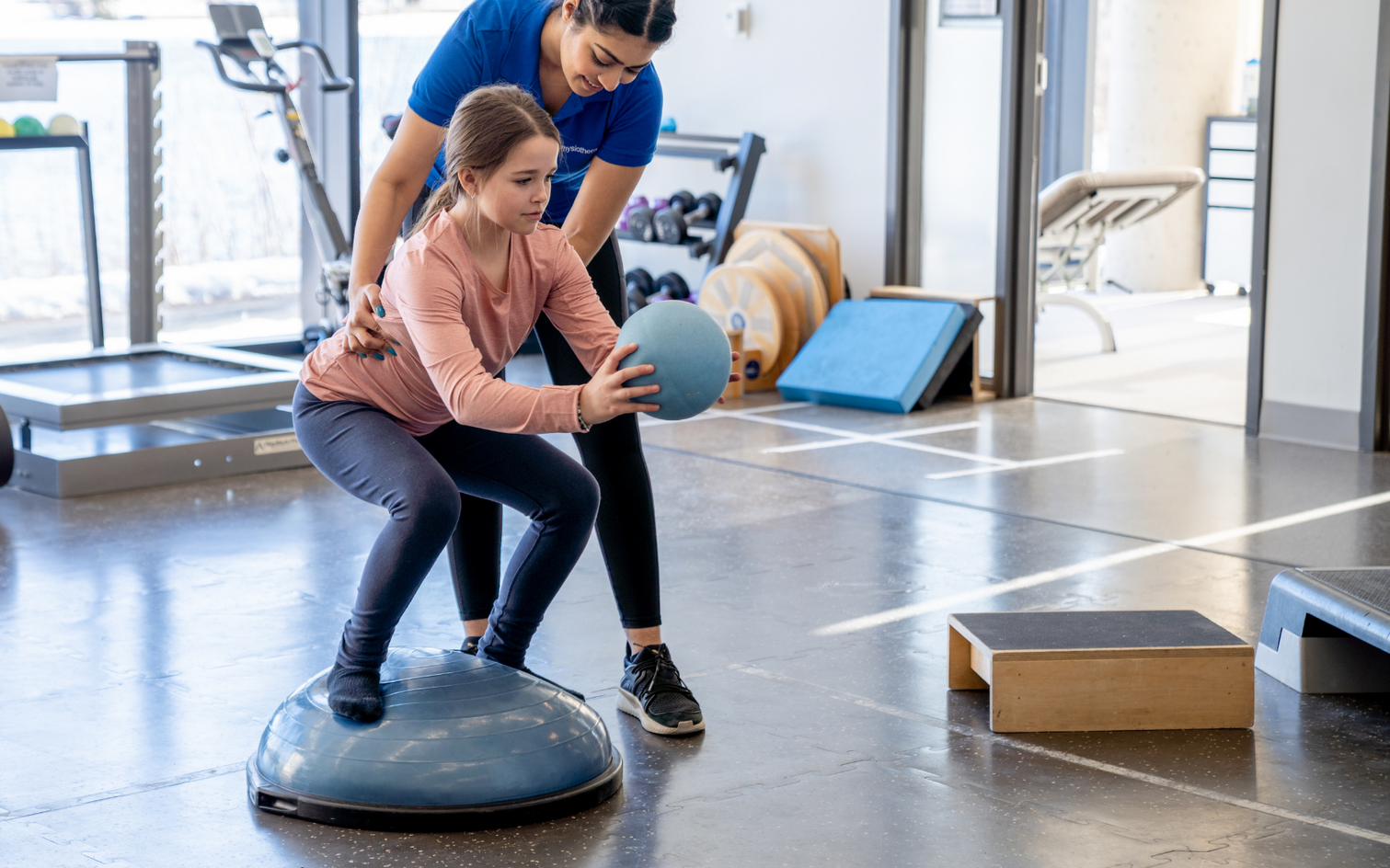
Osgood-Schlatter Disease (OSD), is the most common osteochondritis affecting the lower limb in adolescents playing sport. OSD is a traction apophysitis (apoph-y-si-tis), meaning inflammation (-itis) of the growth area of the bone where a tendon attaches (the apophysis). It is caused by repetitive traction or overload. Activities such soccer, volleyball, basketball, and gymnastics that involve repetitive jumping, sprinting, and change of direction, produce repetitive stress on the growth plate at the top of the shinbone, causing inflammation, pain, and discomfort.
Previously considered to be self-limiting, OSD can result in prolonged absences from sport, resulting in a shift towards risk factor identification and injury prevention.
Anatomy 101.
The main structure involved in OSD is the tibial tuberosity, a bony prominence at the top of the shinbone (tibia), about 3-5cm below the bottom edge of the kneecap, where the patellar tendon connects. This patella tendon attaches the quadriceps muscles to the shinbone via the patella (kneecap), allowing for knee extension and leg stability. In growing adolescents, the tibial tuberosity is the site of a growth plate, making it particularly vulnerable to repeated mechanical traction. Overuse can result in microvascular tears, growth plate fractures, and inflammation, leading to the classic symptoms of OSD of pain, swelling and tenderness on palpation, and reduced functional activity.
Who gets OSD?
OSD primarily affects growing adolescents during the bone maturation phase of growth, typically
between the ages of 8 -13 years in females and 10-15 years in males.
OSD is more common in boys than girls (ratio of 14:1) and is often seen in young athletes who participate in sports that involve running, jumping, and rapid changes in direction. The highest incidence occurs in soccer players, with OSD accounting for about 13% of all knee pathologies in soccer players aged 13-15. Intrinsic factors such as age, growth spurts, morphology, and gender, can contribute to the development of OSD, as well as extrinsic factors like excessive training (including intensity, volume and type), early specialization, inappropriate footwear, and poor biomechanics.
Diagnosing Osgood Schlatter Disease
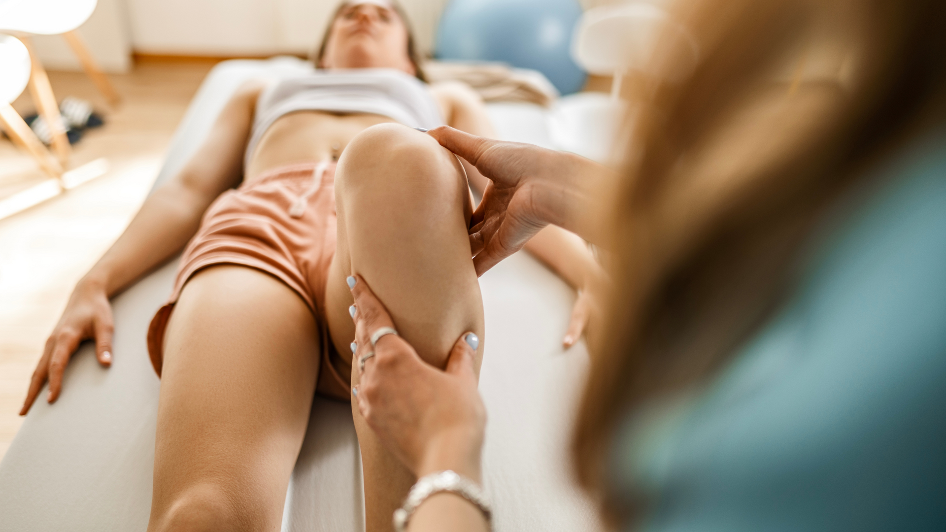
Diagnosis typically involves a thorough interview process and clinical examination by a physiotherapist. Activity history leading up to the development of symptoms and the behaviour of symptoms with different activities provide a good indication of whether OSD is the cause of your symptoms. Importantly, there are several other benign conditions that can cause pain in the same region and have similar symptoms, such as patellofemoral pain syndrome, patellar tendonitis, and Sinding-Larsen-Johansson syndrome, as well as more sinister conditions such as bone tumours that need to be considered in the diagnostic process.
Common signs and symptoms of OSD include pain, often severe enough to result in a limp,
and swelling over the bony prominence below the kneecap.
Pain is often worse during or after physical activity, when descending stairs, kneeling, or after prolonged sitting. Patients may also experience tenderness when palpating the tibial tuberosity and an increase in the size of the tibial tuberosity.
Do I need a scan?
Diagnosis of OSD is clinical and based on the presenting symptoms. However, it is recommended that OSD be confirmed by complementary radiological tests, specifically x-ray, ultrasound, and/or MRI, to allow OSD to be differentiated from other pathologies such as fractures, tumours and tendonitis. X-ray is the first choice, as this helps to identify the severity of OSD and grade involvement. Ultrasound is beneficial for monitoring disease progression due to its low cost and ease of access. MRI (Nuclear MR or NMR) has the highest sensitivity to diagnosing early OSD, however it is associated with high cost and limited access, and so is used sparingly. Your physiotherapist can discuss these investigations with you and refer appropriately where indicated.
Treatment
OSD has long been considered a self-limiting condition however this perspective should be challenged. The affected growth plate tissue, known as bradytrophic tissue, is a component of the cartilage matrix that resists mechanical stress and can take 1-2 years to heal in OSD, a long period of time in a developing adolescent!
A comprehensive treatment program is paramount to educate the patient, accelerate symptom resolution and encourage healing, permit modified exercise to continue, and limit deconditioning and skill degradation.
Education, activity modification and therapeutic exercise are the cornerstone of management of OSD. Reduction of load is critical to allow the inflamed apophysis to settle and commence healing. Once symptoms are controlled, the apophysis can be exposed to graded exercise (strengthening and stretching), and eventual return to sport. This permits the underlying matrix of the apophysis to develop tolerance to mechanical load and long-term tissue resilience.
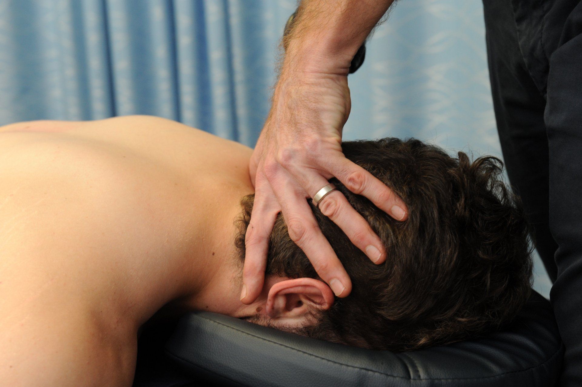
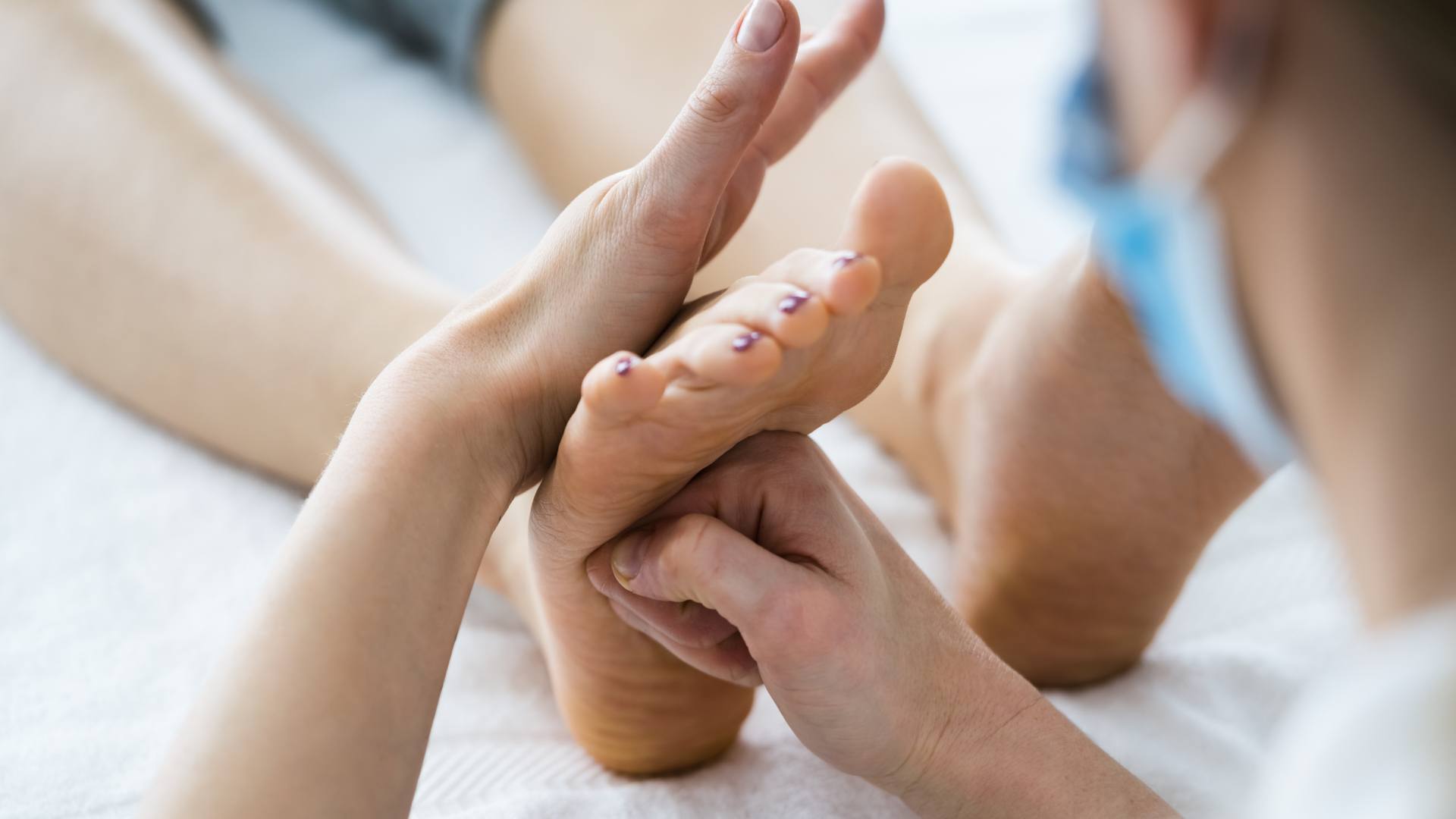
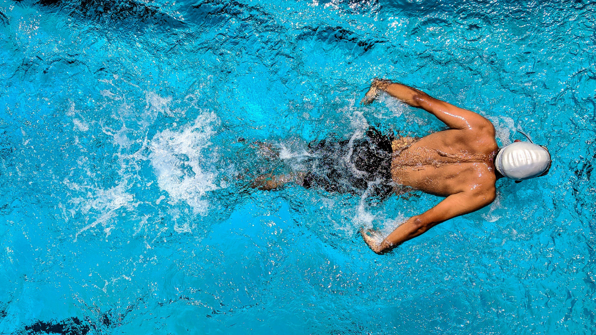
Physiotherapy techniques facilitate this process. Manual therapy, dry needling, taping and bracing can be effective at reducing symptoms, while hydrotherapy and graded land-based exercise programs aimed at improving flexibility, strength, and biomechanics ensure continued exposure to therapeutic load. Pain relief may be prescribed to help manage symptoms, however non-steroidal anti-inflammatory medications (NSAIDs) are discouraged due to their detrimental effects on tendon blood flow and collagen synthesis.
Interventions such as corticosteroid injections, shockwave therapy, and handheld percussive therapies (eg. Theragun), have limited peer reviewed evidence supporting their effectiveness and are not routinely recommended. Surgery is only recommended where conservative treatment has failed and where bone fragments remain after skeletal maturity has occurred.
Remember, no two treatment programs will be alike. Even within OSD, there can be different factors contributing to your presentation. Follow the advice of your physiotherapist, be diligent with your exercises and give it some time. Your symptoms may have developed over a long period of time and will take a similar period to settle and improve.
How long is it going to take?
The prognosis for OSD varies depending on factors such as the severity and duration of symptoms, the age of the patient, and their commitment to treatment. Early intervention, adherence to prescribed exercises, and open communication with healthcare providers can facilitate more effective recovery.
Programs consisting of activity modification, pain monitoring, preogressive strengthening and structured return to sport have been shown to achieve significant improvements in 80% of subjects in 12 weeks, and 90% of cases in 12 months.
Unfortunately, in some cases symptoms will persist until skeletal maturity is reached and the growth plate closes which may take several years.
The Take Home
Osgood-Schlatter Disease is a common and generally self-limiting condition in growing adolescents that can have significant impacts of participation in sport and physical activity if left untreated. In sporting populations the focus should be on risk factor identification and injury prevention. When symptoms do develop, early diagnosis, education, and appropriate physiotherapy intervention is paramount to alleviate symptoms and promote recovery. By working closely with a physiotherapist, young athletes can return to their activities sooner with improved strength, flexibility, and biomechanics, reducing the risk of recurrence.
Got knee pain and want to know the cause? Give us a call.
At Movement for Life Physiotherapy, we can assess and diagnose the cause of your knee pain and let you know whether you have Osgood Schlatter disease, patellofemoral pain syndrome, patella tendinopathy, or if there is something else going on. With a clear diagnosis and tailored management plan, we'll help get you back to the things you love sooner.
Give us a call now or click on BOOK AN APPOINTMENT to book online.
Sources
- Bloom OJ & Mackler L (2004). What is the best treatment for Osgood-Schlatter disease?
- Clark SC, Jones MW, Choudhury RR, & Smith E (1995). Bilateral patellar tendon rupture secondary to repeated local steroid injections. Emergency Medicine Journal. 12(4):300-1.
- Corbi F, Matas S, Álvarez-Herms J, Sitko S, Baiget E, Reverter-Masia J, & López-Laval I (2022) Osgood-Schlatter Disease: Appearance, Diagnosis and Treatment: A Narrative Review. Healthcare. 2022; 10(6):1011. https://doi.org/10.3390/healthcare10061011
- Gholve PA, Scher DM, Khakharia S, Widmann RF, & Green DW (2007). Osgood Schlatter syndrome. Current Opinion in Pediatrics, 19(1), 44-50.
- Kaya DO, & Toprak U (2017). Osgood-Schlatter disease: diagnosis and treatment. EFORT Open Reviews, 2(11), 447-453.
- Neuhaus C, Appenzeller-Herzog C, & Faude O (2021). A systematic review on conservative treatment options for OSGOOD-Schlatter disease. Physical Therapy in Sport, 49, 178-187.
- Pihlajamäki H, & Visuri T (1999). Long-term outcome after surgical treatment of unresolved Osgood-Schlatter disease in young men: surgical technique. The Journal of Bone & Joint Surgery, 81(10), 1426-1432.
- Rathleff MS, Winiarski L, Krommes K, et al. (2020). Activity Modification and Knee Strengthening for Osgood-Schlatter Disease: A Prospective Cohort Study. Orthopaedic Journal of Sports Medicine. 2020;8(4). doi:10.1177/2325967120911106
- Vaishya R, Azizi AT, & Agarwal AK (2016). Osgood-Schlatter disease: A review of literature. Journal of Clinical Orthopaedics and Trauma, 7(4), 244-248.
- Weiner DS, & Macnab I (1971). The "traction" apophysitis. The American Journal of Sports Medicine, 3(3), 152-154.
